For samples of biofluids, especially for urine samples, the normalization is a crucial pre-processing step. The normalization tries to account for different dilutions of samples. Animals drinking different amounts of water excrete urine samples with different dilutions. Thereby the concentrations of most metabolites are dilluted by the sam factor (the so-called overall dilution factor or overall concentration factor). In metabonomics these overall dilution of urine is usually not of interest (although drugs can also influence the dilution of urine). Metabonomic investigations are typically interested in relative changes of concentrations of metabolites. Therefore, the normalization procedure scales urine samples (one scaling factor per sample) to represent the same overall concentration. Usually a so-called integral normalization is applied in metabonomics. Thereby it is assumed, that the area below the spectrum represents the overall concentration of the corresponding sample. Consequently the spectra are scaled to the same integral (usually 1 or 100). Drawbacks of the integral normalization procedure and an improved normalization procedure are demonstrated on the next page.
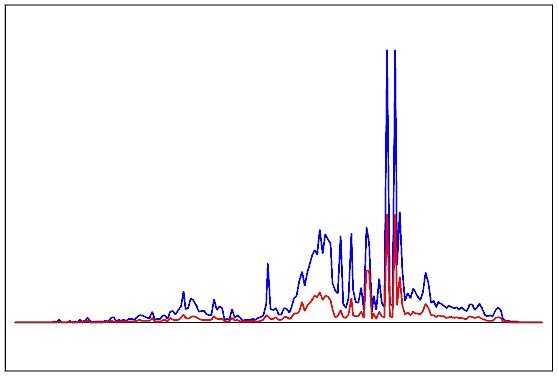
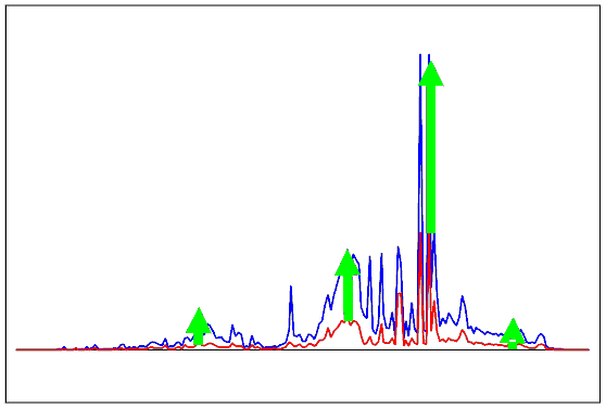
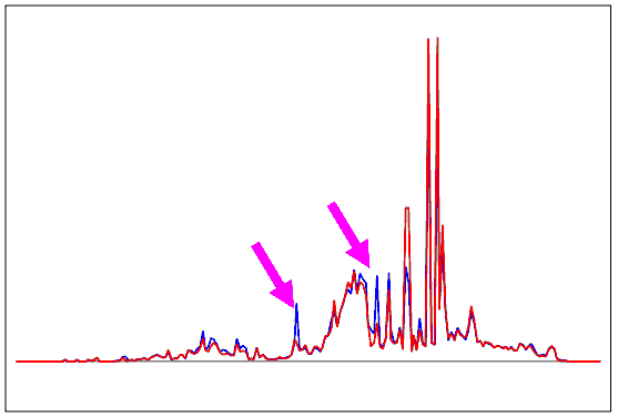
In the figures below, two spectra are shown with different dilutions. The blue spectrum is concentrated by a factor of about 3 resulting in an integral of about three times as high as the red spectrum. Due to the different overall concentrations, both spectra seem to look very different. The integral normalization procedure up-scales the red spectrum by a factor of three to render the areas below the spectrum equal. The third figure shows that besides of the overall dilution both samples are very similar. The two arrows indicate the two parts of the spectra with the most significant differences. These differences are based on relative changes of metabolites and are of interest for metabolic profiling.
NMR spectra of two samples with different dilutions. The sample colored by red is diluted by a factor of three. Therefore most parts of the spectrum are down-scaled by a factor of three. Due to the different dilutions, the spectra seem to look very different.
The integral normalization up-scales the red spectrum by a factor of three to render the areas below the two spectra equal.
The normalized spectra are very similar. The two magenta arrows indicate the two parts of the spectra with the most significant differences. These differences are based on relative changes of metabolites and are of interest for metabolic profiling in contrast to changes due to the overal concentration of urine.